Clinical Example: Aneurysmal Bone Cysts
| Aneurysmal bone
cysts are uncommon benign vascular
bone tumors. Their origin is unknown, but they resemble
intraosseous arteriovenous malformations. They may cause
cortical expansion and thinning, leading to pathologic
fracture. They are more common in females than males, in
young patients (less than 20 years old) and in the lower
extremities, pelvis and spine. The most common treatment
is curettage. One out of five will recur after
curettage. After excision, treatment of the margins with
cryotherapy, phenol or methacrylate may reduce
recurrence. However, if there is structural weakening
from circumferential cortical thinning, cytotoxic
marginal treatment may may be too risky and structural
bone grafting may be required. The following two cases are each an atypical demographic: elderly, male, upper extremity. |
| Click on each image for a larger picture |
|
Case 1
This 75 year old gentleman complained of three months
of wrist pain severe enough to prevent playing golf.
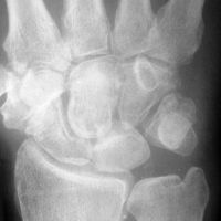
Plain films showed a well circumscribed lobular
lucency in the capitate and STT osteoarthritis. |

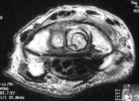
| MRI was
interpreted as inconclusive, differential including an
intraosseous cyst or giant cell tumor with cortical
thinning but no suggestion of malignancy. |


| Dorsal exposure of
the capitate revealed extraosseous extension. |

| The dorsal
capitate was windowed and the tumor was removed with
rongeur and curette. Margins were taken back to
subcortical bone with a high speed burr. Technical point:
The irregular texture of cancellous bone makes it
difficult to visually inspect the margins, and the use
of a small curette only makes this worse. Once the
gross tumor is removed, the margins can be "polished"
with a large high speed burr - the larger the better.
This leaves a a smooth surface and reduces the chance
of residual hidden pockets of tumor. |

| Intraoperative
fluoroscopy of the defect after marginal excision with
a burr... |

| and then after the
defect was filled with corticocancellous iliac crest
bone graft to improve structural stability: |

| Pressure fit bone
graft in place. |

| Pathology:
decalcified specimen showed empty spaces, hemorrhage,
stromal elements and multinuclear osteoclasts - not to
be confused with giant cells. |


| Graft
incorporation at 6 months postop. |

|
Case 2
This elderly gentleman presented with an enlaging
tumor of his thumb tip. He did not know how long
it had been present. He complained that it bled
frequently, was unstable, and he requested an
amputation. He had no lymphadenopathy or evidence of
metastatic disease on chest Xray. |

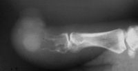
| Plain films showed
loss of the distal 2/3 of the distal phalanx. |

| Excisional biopsy
in the form of an interphalangeal disarticulation: |


| Gross pathology
after decalcification. |

| Microscopic
pathology similar to the prior case, confirming the
diagnosis of aneurysmal bone cyst. |


| Search for... aneurysmal bone cyst
|
Case Examples Index Page | e-Hand home |